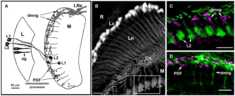
Figure 3.
Anatomical relationships between glial cells (eg, epithelial glial cells of the lamina, dmng, distal medulla neuropil glia) and their putative targets, the L1 and L2 monopolar cells, as well as terminals of some clock neurons (LNs). (A) Diagram showing the relative locations of L1 and L2 in the lamina and distal medulla, the terminals of PDF immunoreactive lateral neurons (LNs), lamina neuropil epithelial glia (eg), and distal medulla neuropil glia (dmng). L, lamina; M, medulla. (B) Morphology of the L2 monopolar cell in the optic lobe of 21D-GAL4 × UAS-S65T-GFP transgenic flies. Cell bodies of L2 are distributed beneath the retina (R), in the region of the lamina cortex (Lc); the axons and dendrites of these cells are in the lamina neuropil (Ln), and their terminals in the medulla (M). Ch, external chiasm. Scale bar: 10 μm. (C,D) The region of the medulla seen as an insert in (B). (C) GFP labeled terminals of L2 and distal medulla neuropil glia (dmng) showing Ebony-like immunoreactivity. Scale bar: 10 μm. (D) The distal medulla neuropil glia (dmng) of Repo-Gal4 × UAS-S65T-GFP transgenic flies and terminals of PDH-immunoreactive LNs (PDF) labeled with anti-PDF antibody. The PDF-positive LNs send projections into the region where the terminals of L2 and Ebony-expressing dmng are located. Scale bar: 10 μm.

