
FIGURE 1.
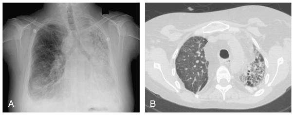
Radiologic images of Patient 6 from this case study, with Fusarium proliferatum, Aspergillus species, and Nocardia asteroides isolated from sputum samples: A, Anteroposterior chest radiograph showing focal consolidation in the mid- to lower right lung likely representing pneumonia following a right thoracotomy and right lung transplantation. There are small bilateral pleural effusions, and left hemithoracic volume loss and interstitial fibrosis or traction bronchiectasis of native left lung. B, Axial CT scan without contrast showing fibrosis in the native left lung as evidenced by the honeycombing architectural distortion. The lower lobes demonstrate ground glass opacity and consolidation. The right transplant lung demonstrates diffuse ground glass opacity and focal consolidation with associated centrilobular nodularity.