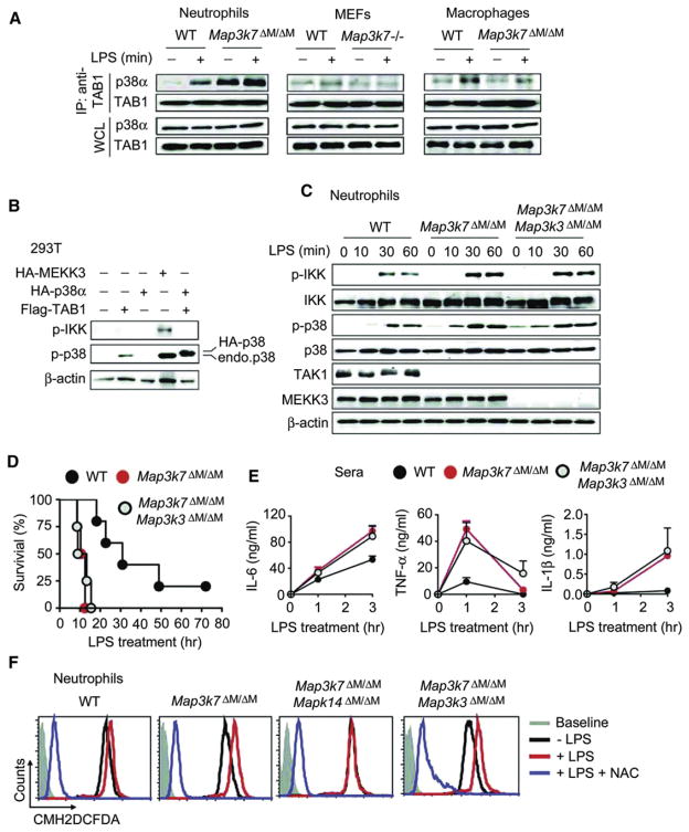
Figure 7. Multiple Pathways Are Involved in p38 Activation in TAK1-Deficient Neutrophils.
(A) Interaction between TAB1 and p38 in neutrophils, peritoneal macrophages, and MEFs. Cells were treated with or without LPS for 15 min and cell lysates were immunoprecipitated (IP) with anti-TAB1 followed by immunoblotting with anti-p38α and anti-TAB1.
(B) 293T cells were transfected with Flag-TAB1, HA-p38α, HA-MEKK3, or empty vector alone, followed by immunoblot analysis of cell lysates with indicated antibodies.
(C) WT, Map3k7ΔM/ΔM, and Map3k7ΔM/ΔMMap3k3ΔM/ΔM neutrophils were treated with LPS, and cell lysates were immunoblotted with indicated antibodies.
(D) Survival of WT (n = 5), Map3k7ΔM/ΔM (n = 4), and Map3k7ΔM/ΔMMap3k3ΔM/ΔM (n = 4) mice treated with high-dose LPS (25 mg/kg). Serum concentrations of IL-6, TNF-α, and IL-1β were measured at 0, 1, and 3 hr after LPS injection.
(E) Serum concentrations of IL-6, TNF-α, and IL-1β were measured at 1 hr and 3 hr after LPS challenge.
(F) WT, Map3k7ΔM/ΔM, Map3k7ΔM/ΔMMapk14ΔM/ΔM, and Map3k7ΔM/ΔMMap3k3ΔM/ΔM neutrophils were pretreated with or without NAC (5 mM) for 30 min followed by LPS stimulation for 3 hr. ROS production was measured by staining cells with CM-H2DCFDA for 30 min followed by flow cytometry.
Results are representative of at least three independent experiments. See also Figure S4.

