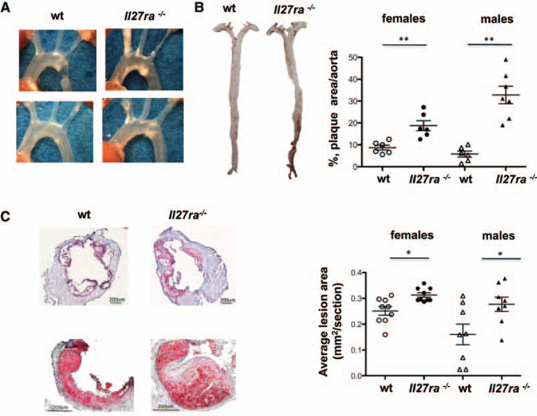
Figure 1. Increased atherosclerotic plaque area inIl27ra−/−bone marrow–transplanted mice.
Ldlr−/− mice were lethally irradiated and reconstituted with 5×10⁶ unfractioned bone marrow cells from C57BL/6 (wild-type [wt]) or Il27ra−/− mice. After 4 weeks of reconstitution, mice were fed with western diet for 16 weeks. A, Images of aortic arch of Ldlr−/− mice receiving Il27ra−/− bone marrow or C57BL/6 (wt) control. B, Representative en face images of Sudan IV–stained whole aortas of Ldlr−/− mice receiving Il27ra−/− or wt bone marrow. B, right, Quantification of plaque area as percentage of aortic surface in Ldlr−/− mice receiving Il27ra−/− or wt bone marrow (n=6–7 females or males per group, respectively). C, left, Representative images of aortic roots (top) and single valve (bottom) of Ldlr−/− mice receiving Il27ra−/− or C57BL/6 (wt) bone marrow. C, right, Aortic root lesions were quantified on frozen sections stained with Oil-red-O in the 300 µm following the aortic valve (wt [n=8–9 males or females] mice and Il27ra−/− [n=8–9 males or females] mice from 3 experiments).

