Figure 3.
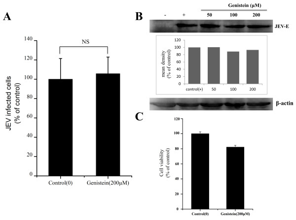
Effects of genistein on JEV infection. (A) PK15 cells were treated with 200 μM genistein for 1 h and infected with JEV in the presence of inhibitor; 36 hours post-infection (hpi), the cells were fixed and stained with anti-JEV E primary and Alexa488 anti-mouse IgG secondary antibodies. The percentage of internalized viruses of drug-treated cells was determined by flow cytometry and normalized to the value for the control. NS, not significant. (B) The cells were treated with increasing concentrations of genistein as indicated, then infected with JEV; 36 hpi, the cells were lysed and processed for western blot analysis of JEV E protein. β-actin was used as an internal loading control. +, control cells untreated with genistein; -, control without JEV infection. The mean densities of the protein bands are shown as a bar graph under the immunoblot. (C) PK15 cells were untreated (control) or treated with genistein (200 μM) for 6 h at 37°C. Cell viability was determined by CCK-8 kit. The results are presented as the mean ± SD.

