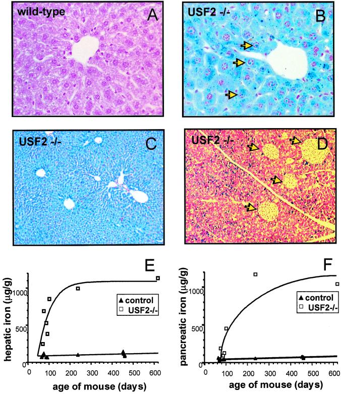
Figure 1.
Iron accumulation in liver and pancreas of Usf2−/− mice. Liver and pancreas were fixed in formaldehyde and stained with the Perls' stain for iron. Nonheme iron stains blue. Liver sections are from an 8-month-old wild-type mice (×50) (A), an 8-month-old Usf2−/− littermate (B), and a 19-month-old Usf2−/− mouse (×10) (C). Pancreas section in D is from an 8-month-old Usf2−/− mouse (×12.5). Arrowheads in C indicate iron in the nucleus of the hepatocyte. Arrowheads in D point to islets of Langerhans scattered throughout the exocrine tissue. (E and F) Age-dependent hepatic and pancreatic nonheme iron concentration (micrograms of iron per gram dry tissue) as measured in control (wild-type and heterozygote mice, ▴) and Usf2−/− mice (□).

