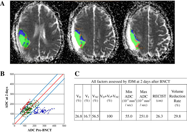
Figure 1.
Representative case: Regions of interests were drawn for tumor image by using anatomical images (A). Red voxels represent areas within the tumor where ADC increased (> 55 × 10-5 mm2/sec); blue voxels represent decreased ADC (< 55 × 10-5 mm2/sec), and green voxels represent no change. These thresholds represent the 95% confidence intervals for change in ADC for the uninvolved cerebral hemisphere (B). VD, VI, VNC, Min/Max ADC, RECIST, and the volume of enhanced lesions on Gd-MRI over time showed 26.8%, 16.7%, 56.5%, 55.0 10-5 mm2/sec, 251.0 10-5 mm2/sec, 26.3 cm, and 29.8% at 2 days after BNCT, respectively (C).

