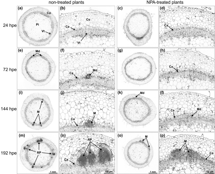
Fig. 4.
Light microscopy of stem base of P. hybrida cuttings during rooting under non-treated and NPA-treated conditions. In all micrographs, semi-thin cross-sections from 1 to 4 mm above the excision site are shown. a, b 24 hpe control cuttings. c, d 24 hpe NPA-treated cuttings. e, f 72 hpe control cuttings. g, h 72 hpe NPA-treated cuttings. i, j 144 hpe control cuttings. k, l 144 hpe NPA-treated cuttings. m, n 192 hpe control cuttings. o, p 192 hpe NPA-treated cuttings. Ca cambium, Co cortex, M meristem of AR, Md meristemoid of AR, Pi pith, RP primordia of AR, Vt vascular tissue, hpe hours post excision

