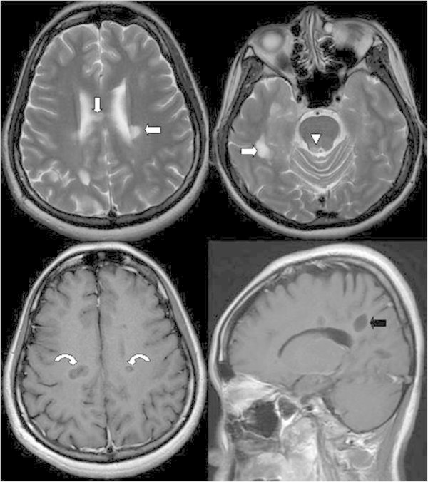
Figure 1.

Hyperintense lesions in the bilateral periventricular white matter (arrow), body of the corpus callosum (arrow), and periaqueductal grey matter (arrowhead) are noted on the T2-weighted image. After gadolinium contrast injection, ring enhancement over these lesions is noted on the T1-weighted image (curved arrows), which is suggestive of active demyelination. In the sagittal view, multiple sclerosis plaques with a typical perpendicular orientation at the callososeptal interface are noted (Dawson’s fingers) (black arrow).
