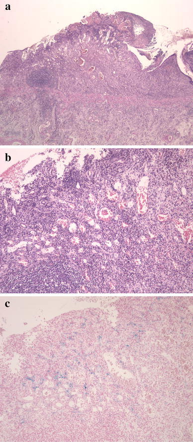
Fig. 6.

Stenotic portions of the ileum formed an ulcer Ul-II excluding the muscularis mucosa. Fibrin and fibrous connective tissue covered the ileal lumen. Inflammatory cell infiltrate, particularly of lymphocytes and eosinophils, was observed throughout all layers. Dilatation and congestion of the submucosal capillary vessels were observed (H&E staining: a low-power field, b high-power field). Haemosiderin staining revealed sideroferous cells in the submucosal layers (c)
