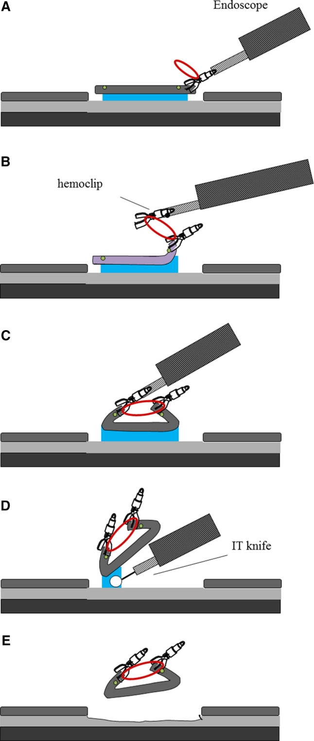
Fig. 3.

A Marking dots are made on the circumference of the target tumor, outlining the margin of the lesion. After injection of a saline solution into the submucosa, the tumor is separated from the surrounding normal mucosa by complete incision around the lesion using the IT knife. B, C The device connects to the edge of the exfoliated mucosa and the opposite side of the lesion. D In pulling the lesion up and opening the resection margin, dissection can be rapidly accomplished by tension from the elastic material. E After dissection, the device is recoverable with the lesion. Device can be easily removed from the lesion with forceps
