
Figure 1. β-AR enhances ICa,L in mouse ventricular cardiomyocytes via the activation of ACVI.
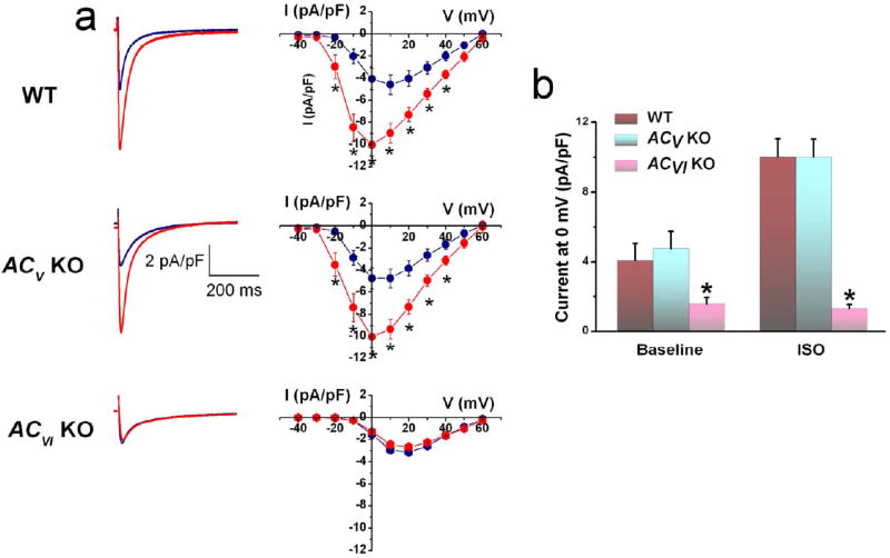
(a) Representative ICa,L recorded from ventricular myocytes isolated from WT, ACV KO, and ACVI KO, respectively. Current traces were elicited using a voltage step of 0 mV for 500 ms from a holding potential of -55 mV at baseline (blue) and after 20 minutes of ISO (red). The corresponding currentvoltage (IV) relations elicited using a family of voltage steps from -40 to +60 mV from a holding potential of -55 mV are shown to the right. (b) Schematic representation of the known regulation of ICa,L by β-AR stimulation. (c) Summary data of ICa,L density (in pA/pF) from the three groups of mice (n=8 for each group, *P<0.05).