
Figure 2. ACV isoform is critical for β2AR enhancement of ICa,L in the mouse ventricular myocytes.
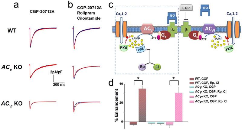
(a) Representative ICa,L recorded from ventricular myocytes isolated from WT, ACV KO, and ACVI KO, respectively. Current traces were elicited using a voltage step of 0 mV for 500 ms from a holding potential of -55 mV at baseline (blue) and after 20 minutes of ISO (red). β1ARs were blocked by CGP-20712A (CGP). No significant effect of β2AR stimulation was observed. (b) Application of a PDE3 blocker (rolipram, Rp) and a PDE4 blocker (cilostamide, CI) revealed the effect of β2AR stimulation only in WT and ACVI KO animals. (c) Schematic representation of the experimental results suggesting that ACV is localized within the same compartment as β2ARs and PDE shown in the box as outlined, separated from β1ARs. (d) Summary data of the percentages of ICa,L enhancement by β2AR stimulation in the three groups of mice with and without PDE blockers (n=7-8 for each group, *P<0.05).