
Figure 3. β1ARs mediate the enhancement effects on ICa,L via both ACV and ACVI isoforms.
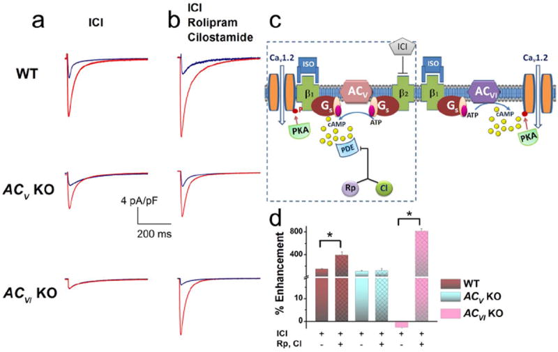
(a) Representative ICa,L recorded from ventricular myocytes isolated from WT, ACV KO, and ACVI KO, respectively. Current traces were elicited using a voltage step of 0 mV for 500 ms from a holding potential of -55 mV at baseline (blue) and after 20 minutes of ISO (red). β2-ARs were blocked by ICI- 118,551 (ICI). No β1AR stimulation of the ICa,L was observed in the ACVI KO group. (b) Application of a PDE3 blocker (Rp) and a PDE4 blocker (CI) revealed the stimulatory effect of β1AR in the ACVI KO mice and further enhanced the effects of β1AR in the WT mice. (c) Schematic representation of the experimental results which suggest that β1ARs mediate the enhancement effects on ICa,L via both ACV and ACVI isoforms. Moreover, only the effect through the ACV isoform is restricted by PDE. (d) Summary data of the percentages of ICa,L enhancement by β1AR stimulation in the three groups of mice with and without PDE blockers (n=6-8 for each group, *P<0.05).