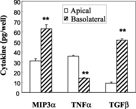
FIG. 1.
Constitutive release of MIP3α, TGF-β, and TNF-α by polarized uterine epithelial cells. Rat uterine epithelial cells were grown to confluence for 5 to 6 days in F12K complete medium at 37°C in NUNC cell culture inserts. Following a 24-h incubation in fresh medium, apical and basolateral media were collected for analysis of MIP3α and TNF-α by ELISA or of TGF-β by bioassay. Values shown are mean cytokine levels ± standard errors from five or more wells per group. Results for each cytokine are representative of three or more separate experiments. **, significantly (P < 0.01) different from cytokine level measured in the apical chamber.

