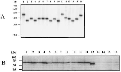
FIG. 4.
Southern and Western blot analyses of H. ducreyi strains to detect lspB genes and LspB protein expression. (A) For Southern blot analysis, chromosomal DNA from each strain was digested with EcoRV, electrophoresed through a 0.8% (wt/vol) agarose gel, transferred to nitrocellulose, and probed with an α-32P-labeled 1.37-kb DNA region of the H. ducreyi lspB ORF. The positions of DNA size markers are indicated on the left. (B) Whole-cell lysates of H. ducreyi strains were subjected to SDS-PAGE and Western blot analysis by using a polyclonal LspB antiserum as described in Materials and Methods. The positions of molecular size markers are indicated on the left. Lane 1, 35000; lane 2, RO18; lane 3, 181; lane 4, CA173; lane 5, WPB506; lane 6, BG411; lane 7, 041; lane 8, 1145; lane 9, 1151; lane 10, Cha-I; lane 11, Hd12; lane 12, CIP 542; lane 13, A77; lane 14, 6V; lane 15, E1673; lane 16, 78226.

