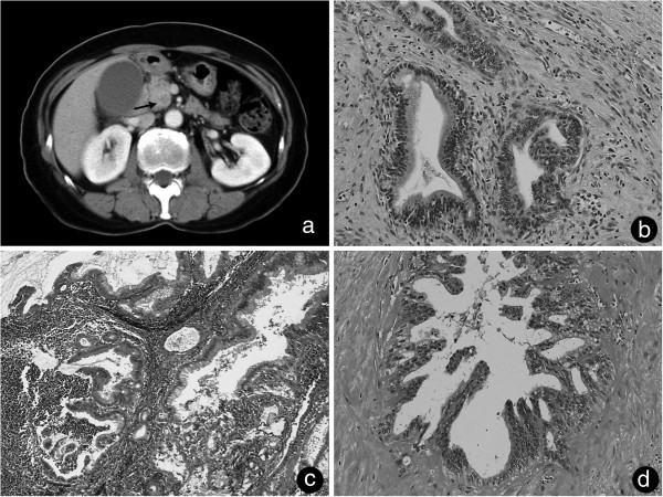
Figure 1.
Primitive tumor CT imaging and histopathological findings. (a) Computed tomography on admission showed a 22 mm × 25 mm low-density mass around the uncus of the pancreas (arrow). (b) Microscope analysis showed a well-differentiated tubular adenocarcinoma surrounded by an extracellular matrix (hematoxylin and eosin, ×40). (c) The para-aortic lymph node had a well-differentiated tubular adenocarcinoma. (d) Resected perineural tissue with a histologic structure similar to the primitive tumor with hyalinization.

