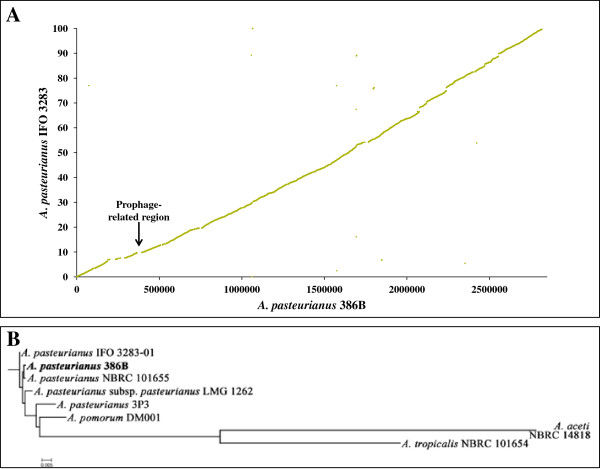
Figure 2.
Comparative analysis of the Acetobacter pasteurianus 386B genome. (A) Synteny plot between the chromosomes of A. pasteurianus 386B and A. pasteurianus IFO 3283. Each dot represents a predicted A. pasteurianus 386B protein having an ortholog in the A. pasteurianus IFO 3283 chromosome, with coordinates corresponding to the position of the respective coding region in each genome. The position of the putative prophage is marked with a vertical black arrow. (B) Phylogenetic tree based on all core genes of the strains included. Multiple sequence alignments of concatenated core gene sequences were calculated within EDGAR. Plasmid sequences were included.

