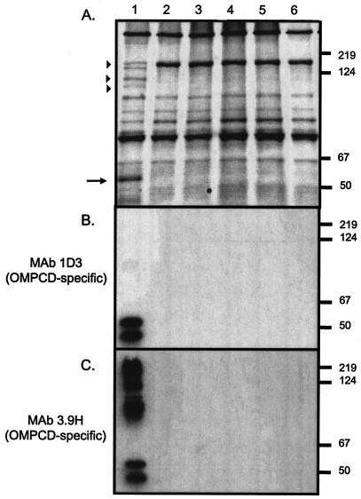
FIG. 4.
Western blot analysis of M. catarrhalis strains. Proteins present in outer membrane vesicles prepared from the wild-type strain O35E (lane 1) as well as the transposon mutants O35E.TN52 (lane 2), O35E.TN313 (lane3), O35E.TN593 (lane 4), and O35E.TN649 (lane 5) and from the isogenic mutant O35E.CD1 (lane 6) were resolved by SDS-PAGE and stained with Coomassie blue (A) or analyzed by Western blotting with the OMPCD-specific MAbs 1D3 (B) and 3.9H (C). Positions of molecular mass markers are shown to the right in kilodaltons. The arrow as well as arrowheads indicate antigens that are missing in the transposon as well as the O35E.CD1 mutants.

