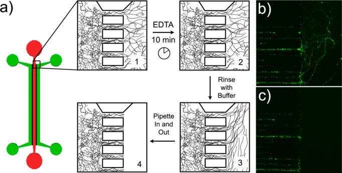
Figure 2.

Samples of pure axonal material can be mechanically isolated using the axonal sample isolation device. (a) Axonal sample isolation device and method. Step 1: Plate neurons in the somal (red) channels and wait until 14 d.i.v. for complete axonal growth. Step 2: Fill axonal (green) channel with EDTA to deadhere axons from coverglass. After 10 min, remove the EDTA and rinse the axons with sample buffer. Step 3: Using fresh sample buffer, pipet in and out at the entrance to the channel to pull and break axons. Step 4: Rinse axonal channels with sample buffer to remove any remaining axonal material. Dendrites in the small connecting channels remain undisturbed. (b) EGFP expressing axons in the connecting channels and axonal chamber prior to and (c) just after removal. Connecting channels are 10 μm wide and spaced 20 μm apart.
