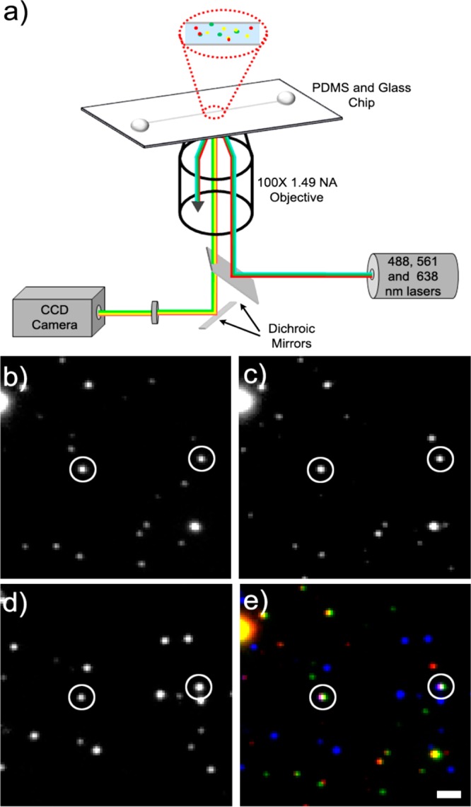
Figure 4.

Fluorescently labeled axonal carrier samples can be viewed in three colors using TIRF microscopy. (a) TIRF imaging setup schematic. (b) Alexa Fluor 488 puncta (p38 rabbit polyclonal primary antibody), (c) Alexa Fluor 568 puncta (p38 rabbit polyclonal primary antibody), and (d) Alexa Fluor 647 puncta (KIF1A mouse monoclonal primary antibody) are (e) checked for colocalization (Alexa Fluor 488 shown in green, Alexa Fluor 568 shown in red, and Alexa Fluor 647 shown in blue). Puncta that fit the three-color colocalization criteria are circled in white. Scale bar is 1.6 μm.
