Figure 1.
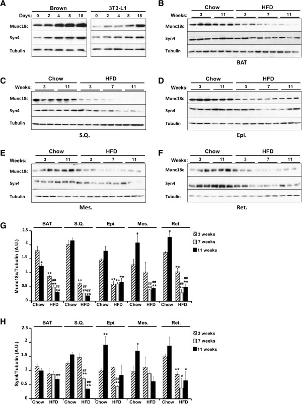
Munc18c expression in adipocytes and adipose tissue depots. A) Immunoblots of Munc18c, syntaxin4 and Tubulin in lysates of brown and white (3T3-L1) adipocytes at different stages of differentiation. B-F) Immunoblots of Munc18c, syntaxin4 and Tubulin in lysates of brown (BAT), subcutaneous (S.Q.), epididymal (Epi.), mesenchymal (Mes.), and retroperitoneal (Ret.) adipose depots of mice fed regular chow or HFD for the indicated times. Each lane represents a sample from a separate animal. Bar graphs represent normalized data for Munc18c (G) and syntaxin4 (H) expression normalized to Tubulin and presented as means ± SEM. (*; P ≤ 0.05, **; P ≤ 0.01) indicate significant difference, in each adipose depot, between all groups versus mice fed regular chow for 3 weeks, and (#; P ≤ 0.05, ##; P ≤ 0.01) indicate significant difference between mice fed HFD for 7 and 11 weeks versus mice fed HFD for 3 weeks.

