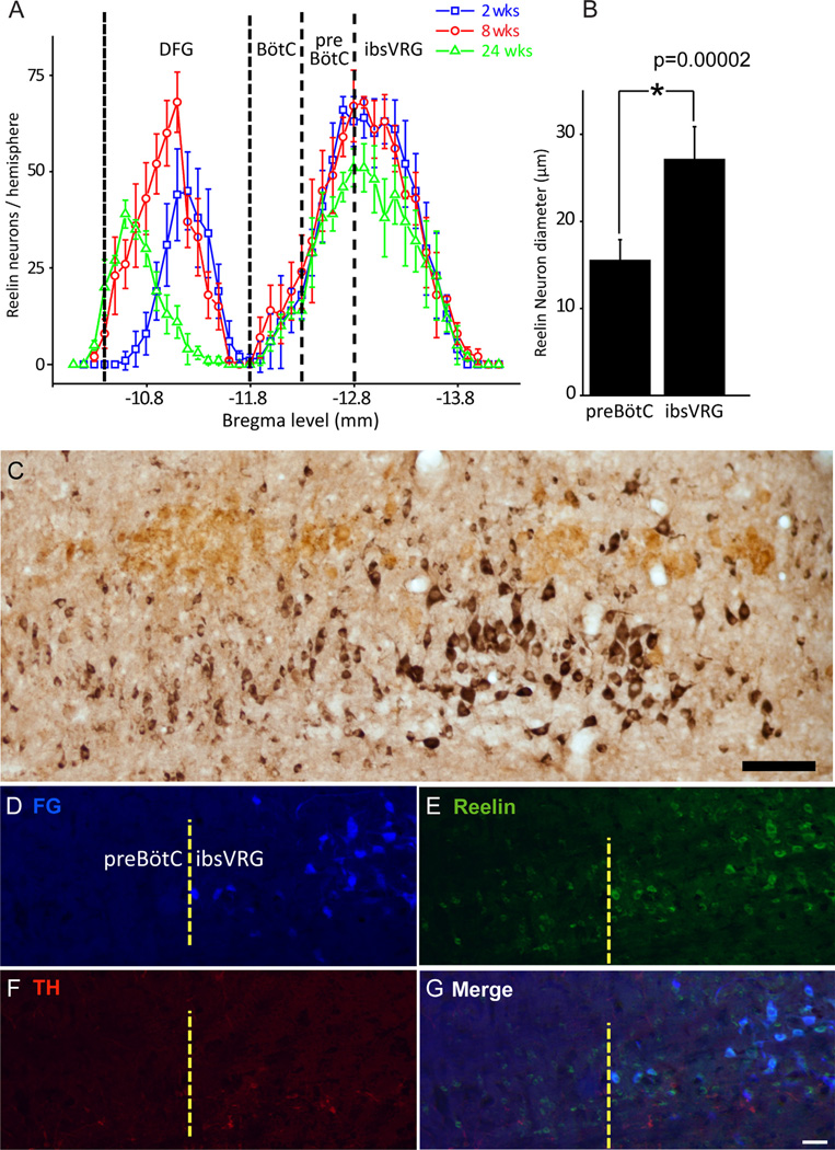
Figure 5.
Distinctive reelin populations set the anatomic boundary of pre-BötC and ibsVRG. A: Reelin neuron rostrocaudal distibution in ventrolateral medulla at three different ages. One reelin neuron group resides in the region from Bregma levels −10.4 to −11.7 mm in DFG; another reelin neuron group located in pre-BötC and ibsVRG with peak cell numbers from Bregma levels −12.7 to −12.9 mm. Caudal edge of the facial nucleus: Bregma −11.7 mm. B: Pre-BötC reelin neurons have significantly smaller soma diameter than those of ibsVRG (ANOVA, P = 0.00002). C: Sagittal view depicting pre-BötC and ibsVRG reelin populations, labeled for reelin (black) and ChAT (brown). Double label with ChAT was used to reveal the motor neurons in the sections. D–G: ibsVRG neurons retrogradely labeled from the C2–C4 level of the spinal cord with FG (D) were stained for reelin (E) and TH (F). G: Merge. Note that FG-positive cells in ibsVRG are colocalized with reelin but do not express TH. ChAT, choline acetyltransferase; FG, Fluorogold; ibsVRG, inspiratory bulbospinal ventral respiratory group; TH, tyrosine hydroxylase. Scale bars = 100 µm in C; 50 µm in G (applies to D–G). [Color figure can be viewed in the online issue, which is available at wileyonlinelibrary.com.]

