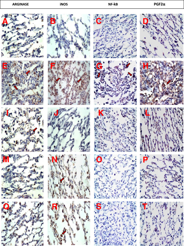
Figure 8.
Guinea pig lung tissue samples obtained from controls (panels A, B, C and D), OVA-exposed and vehicle-treated animals (panels E, F, G and H), OVA-exposed and nor-NOHA treated animals (panels I, J, K and L), OVA-exposed and 1400 W treated animals (panels M, N, O and P), and OVA-exposed and nor-NOHA and 1400 W treated animals (panels Q, R, S and T). Immunohistochemistry analysis for arginase (panels A, E, I, M and Q - ×400), iNOS positive cells (panels B, F, J, N and R - ×1000), NF-kB (panels C, G, K, O and S – ×400), and PGF2α (panels D, H, L, P and T - ×400). The control group showed low amounts of iNOS positive cells, NF-kB, arginase and PGF2α in alveolar tissue sections. In contrast, the distal lung parenchyma of OVA-exposed and vehicle-treated animals showed an increase in the amount of arginase, iNOS-positive cells, NF-kB and PGF2α. The 1400 W and nor-NOR treatments in ovalbumin-exposed animals reduced all these parameters compared to the OVA group. Both treatments in conjunction contributed to a greater PGF2α reduction.

