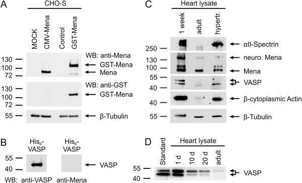
Figure 1.
Stage-dependent expression of cytoskeletal proteins in the mouse heart. (A, B) Characterization of Mena-specific antibodies. (A) CHO-S cells, either transiently transfected with murine Mena (CMV-Mena) or stably transfected with GST-tagged Mena (GST-Mena), were lysed and analyzed by Western blotting with Mena-specific (upper panel) or GST-specific (middle panel) antibodies. As control, lysates of MOCK-transfected cells or lysates of CHO-S cells without stable integration of GST-Mena were run on the same gels. β-Tubulin served as loading control (lower panel). (B) 100 ng purified VASP protein was probed by Western blotting with anti-VASP (left panel) and anti-Mena (right panel) antibodies. (C, D) Stage-dependent expression of cytoskeletal proteins in the mouse heart. (C) Lysates of 1-week-old, adult, and hypertrophic adult mouse hearts were probed by Western blotting with antibodies against αII-Spectrin, Mena, VASP, and β-cytoplasmic actin. Blotting for β-Tubulin was used as invariant loading control. Two Mena isoforms are expressed in the mouse heart, the general (~80 kDa) and the neuronal-specific (~140 kDa) form. Depending on its phosphorylation state, VASP migrates at 46 or 50 kDa. (D) VASP expression in the mouse heart at postnatal day 1, 10, and 20 and in the adult heart.

