Figure 4.
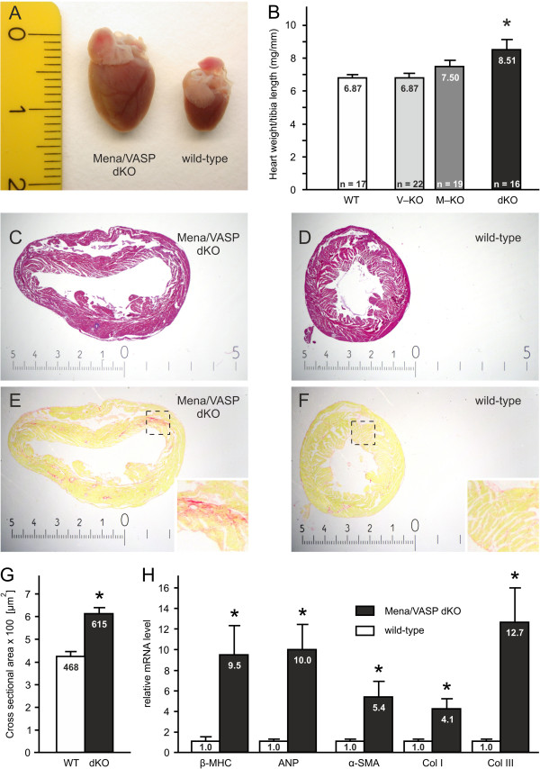
Enlarged and dilated hearts in MenaGT/GTVASP−/− mice. MenaGT/GTVASP−/− (dKO) animals display macroscopically enlarged hearts (A, photography, scale in cm) and significantly increased heart weight to tibia length ratios as compared to wild-type (WT) controls (B; ANOVA, *P<0.05). (C, D) Hematoxilin and eosin stained cross sections of MenaGT/GTVASP−/−(C) and wild-type (D) hearts, demonstrating dilated ventricles in the double-deficient animals. (E, F) Picrosirius red stained heart cross sections of dKO (E) and WT (F) mice to visualize collagen fibers/interstitial fibrosis. Magnified views of the indicated areas are shown as insets. The scales in images C-F are given in mm. (G) Cross sectional areas of cardiomyocytes from WT and MenaGT/GTVASP−/− hearts quantified by morphometric analyses (five animals per group, 25 cardiomyocytes per animal, *P<0.05). (H) Quantitative real-time RT-PCR revealed increased mRNA levels of cardiac hypertrophy markers (β-myosin heavy chain, β-MHC; atrial natriuretic peptide, ANP) and fibrosis markers (α-smooth muscle actin, α-SMA; collagen I, Col I; and collagen III, Col III; *P<0.05; n=6) in dKO mice vs. WT controls.

