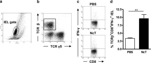
Figure 2.

Increased expression of IFN-γ by TCRβ+CD8+ IEL in N. caninum infected mice. Representative dot plots of (a) cells isolated from the small intestines of C57BL/6 mice (IEL were gated as shown); (b) TCRγδ and TCRβ IEL. (c) Gated TCRβ CD8+ IEL expressing IFN-γ in control (PBS) and N. caninum-infected mice (NcT). (d) Frequency of TCRβ+CD8+ IFN-γ+ IEL in control and N. caninum i.g.-infected mice, 48 h after challenge. Bars represent mean plus one standard deviation of three animals in the PBS group and four animals in the infected mice group. This is one representative result of three independent experiments (*P<0.05).
