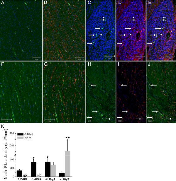
Figure 1.
Neurofilament-M and GAP43 expression by nestin(+) cells in the heart of sham and 1 week post-MI rat hearts. (Panels A &B) In the left ventricle of sham rats, neurofilament-M(+) (green fluorescence) and nestin(+) (red fluorescence) fibres were detected and co-expression of the intermediate filament proteins was not observed. (Panels C, D &E) In the peri-infarct/infarct region of a 4-day post-MI rat heart, innervating neurofilament-M(+) fibres were detected and the preponderance physically associated with nestin(+) fibres (indicated by arrow). Neurofilament-M(+) fibres lacking nestin co-expression were also identified (indicated by arrowhead). (Panels F &G) GAP43(+) (green fluorescence) and nestin(+) (red fluorescence) fibres were detected in the normal rat heart and a paucity co-expressed both proteins. (Panels H, I & J) GAP43/nestin(+) co-expressing fibres (indicated by arrow) were detected innervating the peri-infarct/infarct region 24 hrs after complete coronary artery ligation. The nucleus was identified with TO-PRO-3 staining (blue fluorescence). (Panel K) The density of GAP43/nestin(+) fibres innervating the peri-infarct/infarct region was increased 24 hrs after myocardial infarction and preceded the appearance of neurofilament-M in nestin(+) cells. With ongoing scar formation/healing, the density of GAP43/nestin(+) fibres progressively decreased whereas a concomitant increase in neurofilament-M/nestin(+) fibre density was apparent 4 and 7 days after myocardial infarction. (*) denotes P < 0.05 versus sham, (**) P < 0.05 versus 4 day infarcted rat hearts and (ND) not detected.

