Figure 1.

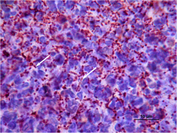
Histological observations of mice tissue infected byN. cyriacigeorgicaGUH-2. Photograph illustrating the immunohistochemistry analysis of kidney cells from a case of fatal septicemia; white arrows indicate filamentous bacteria.

Histological observations of mice tissue infected byN. cyriacigeorgicaGUH-2. Photograph illustrating the immunohistochemistry analysis of kidney cells from a case of fatal septicemia; white arrows indicate filamentous bacteria.