
Figure 5.
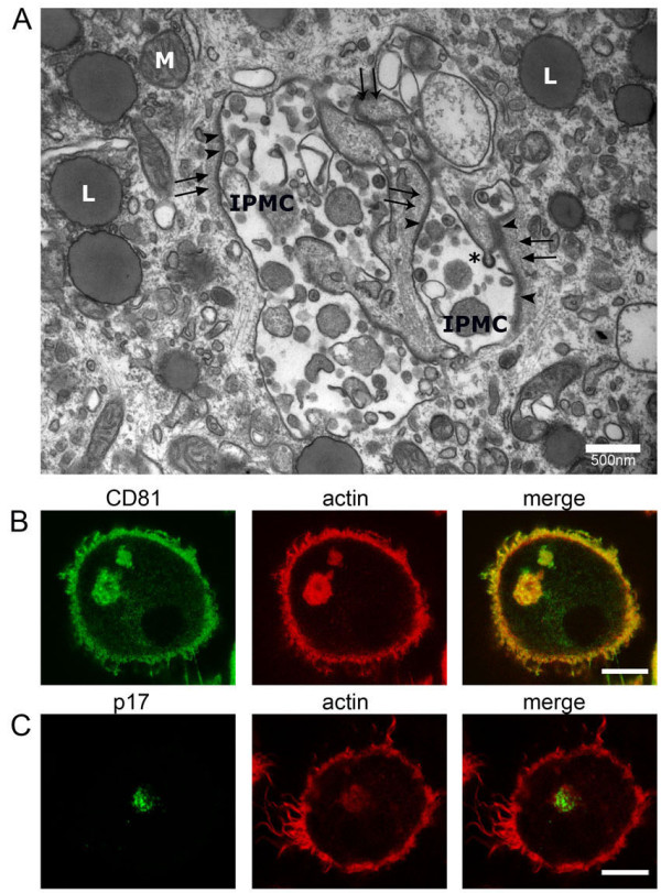
Association of actin with IPMCs. (A) HIV-infected MDMs were embedded in Epon, sectioned and analyzed by transmission EM, as previously described [14]. HIV particles are seen in a complex IPMC. Some of the IPMC membranes are covered with a coat of electron-dense focal adhesion proteins (arrowheads) and a layer of fine filaments (black arrows). L, lipid droplets; M, mitochondrion; the asterisk marks a budding HIV particle. Epon EM was conducted on MDMs from two donors. Scale bar = 500 nm. (B) Uninfected MDMs were stained for CD81 and actin (Alexa Fluor 594-conjugated phalloidin). (C) HIV-1-infected MDMs were stained for the matrix protein p17 and actin. The images show single optical sections acquired with a Leica SPE confocal microscope as above. Scale bars = 10 μm.