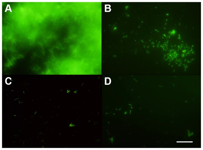
Figure 2. Fluorescence labeling of X. fastidiosa biofilm.

Cells in biofilm were either not treated with NAC (A) or exposed to different NAC concentrations, 1.0 mg/mL (B), 2.0 mg/mL (C), and 6.0 mg/mL (D) and stained with Syto 9 to evaluate the characteristics of the formed biofilm (Scale bar, 20 µm).
