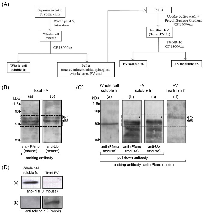
Figure 1. Analysis of P . yoelii food vacuole (FV) associated enolase variants using antibody pull down assays and western blots.
(A) P . yoelii cell fractionation scheme. Various fractions used for analysis are marked in bold. Proteins were separated on 10% SDS PAGE.
(B) Western blot of FV proteome probed with (a) anti-rPfeno (mouse) and anti-Ub (mouse) antibodies. Note that three variants of enolase were observed while two of these (65 & 75 kDa) are ubiquitinated (dotted line box). Matched amounts of proteins were loaded in both the lanes.
(C) Antibody pull-down assays for different sub-cellular fractions using anti-rPfeno (mouse) and anti-Ub antibodies (mouse). Blots of pull-down proteins were probed with anti-rPfeno (rabbit) to detect the presence of enolase.
(D) Western blots of whole cell soluble fraction and total FV probed with ant-PfP0 (cytosolic marker) and anti-falcipain-2 (FV marker) antibodies. Equivalent amount of total proteins were loaded in each lane. Note the enrichment of falcipain-2 and near absence of P0 in FV preparation indicating that preparation used in above experiments is highly enriched in FV.

