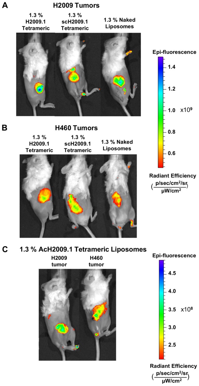
Figure 3. Targeted H2009.1 and control liposomes accumulate in tumors to the same extent.

Subcutaneous αvβ6-positive H2009 (panel A) or αvβ6-negative H460 tumors (panel B) were established in the right flank of NOD/SCID mice. Tumor bearing mice were injected via tail vein with either 1.3% H2009.1 tetrameric, AcH2009.1 tetrameric, scH2009.1 tetrameric, or naked liposomes labeled with the near infrared dye DiR. Animals were imaged at 24, 48, and 72 hours post-liposome injection. Shown are representative images from the 72 hour time point. Despite the αvβ6-targeting abilities of the H2009.1 peptide, all liposomes, except for the AcH2009.1 tetrameric liposomes, accumulated at similar levels in αvβ6-positive H2009 tumors and αvβ6-negative H460 tumors. (C) 1.3% AcH2009.1 tetrameric liposome accumulation in both H2009 and H460 tumors. Note the difference in the epi-fluorescence scale compared to panels A and B.
