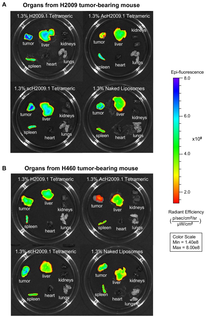
Figure 4. Targeted H2009.1 and control liposomes accumulate in tumors and clear through the liver and spleen.
Subcutaneous αvβ6-positive H2009 or αvβ6-negative H460 tumors were established in the right flank of NOD/SCID mice. Tumor bearing mice were injected via tail vein with either 1.3% H2009.1 tetrameric, AcH2009.1 tetrameric, scH2009.1 tetrameric, or naked liposomes labeled with the near infrared dye DiR. At 72 hours post-injection, the mice were sacrificed and the tumors and organs removed for ex vivo fluorescent imaging. (A) Representative images of liposome accumulation in tumors and organs from αvβ6-positive H2009 tumor bearing mice. Like the in vivo imaging in Figure 3, despite the αvβ6-targeting abilities of the H2009.1 peptide, all liposome formulations except for the AcH2009.1 tetrameric liposomes accumulated in tumors to the same extent. (B) Representative images of liposome accumulation in tumors and organs from αvβ6-negative H460 tumor bearing mice. Similar to the in vivo imaging in Figure 3, the H2009.1 tetrameric and scH2009.1 tetrameric liposomes accumulated in tumors to the same extent, with the AcH2009.1 and naked liposomes accumulating in tumors at levels that are 2-fold lower.

