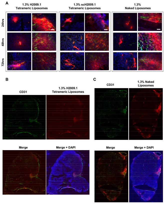
Figure 5. Both targeted H2009.1 and control liposomes accumulate only in the perivasculature regions of H2009 tumors.
Subcutaneous αvβ6-positive H2009 tumors were established in the flank of NOD/SCID mice. Tumor bearing mice were injected via tail vein with either 1.3% H2009.1 tetrameric, scH2009.1 tetrameric, or naked liposomes labeled with the dye DiI. At 24, 48, or 72 hours post-liposome injection, the mice were sacrificed and the tumors removed for sectioning and fluorescent microscopy. Blue – DAPI, Red – DiI-labeled liposomes, and Green – CD31 vasculature stain. (A) 10X images of liposome accumulation in tumors. The white scale bars indicate 100 μm. At each time point, all liposomes are clustered in the areas immediately adjacent to the vasculature, with the same pattern of accumulation for all of the different liposome formulations. Although there are areas of high liposome accumulation, they only occur in vascular-rich areas with large blood vessels. (B) Representative whole tumor image from a mouse injected with 1.3% H2009.1 tetrameric liposomes and sacrificed 48 hours after injection. The liposome accumulation overlaps with the highly vascularized periphery of the tumor. (C) Representative whole tumor image from a mouse injected with 1.3% naked liposomes and sacrificed 48 hours after injection. Like the targeting H2009.1 tetrameric liposomes, the naked control liposomes display the same overlap with the highly vascularized periphery of the tumor.

