
Figure 1. Antiviral effect of IFN-α and RBV combination treatment using a sub-genomic replicon cell line (S3-GFP) and HCV infected Huh-7.5 cells.
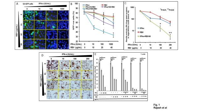
(A) S3-GFP cells were treated with IFN-α and RBV for 72 hours. GFP expression was examined under a fluorescence microscope. (B) GFP positive cells were quantified by flow cytometric analysis. (C) Infected Huh-7.5 cells were treated with IFN-α alone, RBV alone and combination for 72 hours. Renilla Luciferase activity of infected cells was measured and normalized with 1µg of cellular protein. (D) Expression of HCV core protein was measured by immunostaining and (E) core positive cells in five different high power fields (hpf) at 40X magnification were counted under a light microscope. Quantitative assessment of the number of HCV positive cells with mean and standard deviation of the combination treatment are compared.