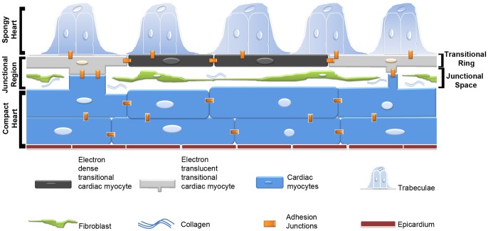
Figure 8. Schematic representation of the interface region between compact and spongy myocardium in the adult zebrafish.
The interface consists of a complex junctional region with an interstitial space populated by a fibroblast network and collagen fibrils. A ring of flattened cardiac myocytes forms the base of the projecting luminal trabeculae, and at discrete intervals make contacts with the compact myocardium though bridges across the junctional space. These areas of contact display complex adhesions junctions including desmosomes and fascia adherents that integrate the two myocardia into a structural unit and may mediate electrical and functional integration of the two myocardia. CCM, compact cardiac myocytes; ETTCM, electron translucent transitional cardiac myocytes; EDTCM, electron dense transitional cardiac myocytes.

