
Figure 1.
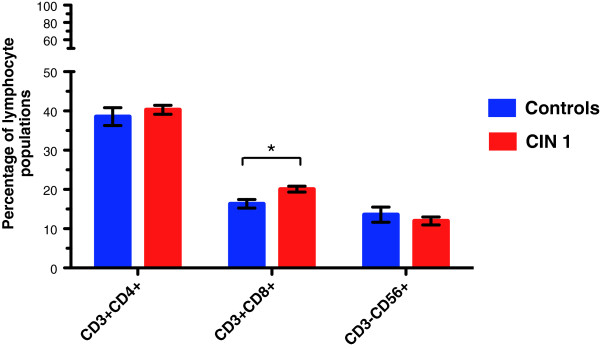
CD8+ T cells are significantly increased in CIN 1 patients. Flow cytometric analysis of peripheral blood in CIN 1 patients or normal donors was performed in order to evaluate variations in the percentages of T cells and NK cells. Gate was drawn around the lymphocyte population, which was then analyzed separately with the corresponding antibodies (CD3+ FITC/CD4+ PECy5, CD3+ FITC/CD8+ PECy5, and CD3- FITC/CD56+ PECy5 for detection of CD4 T cells, CD8 T cells and NK cells, respectively). Analysis from the lymphocyte gate shows that both CD4+ T cells and CD8+ T cells were increased in CIN 1 patients, while NK cells were diminished; however, we only observed a significant increase in the CD8+ T cell population. Statistical analysis was performed through Mann–Whitney U test and data were expressed as mean ± SEM; *p = 0.015.