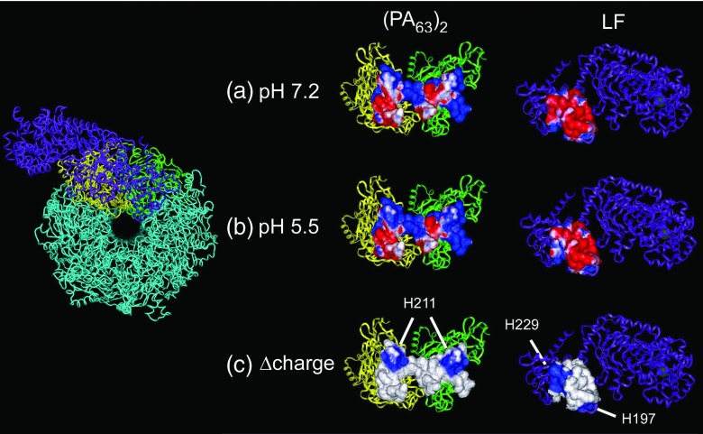
Figure 6.
Hypothetical pH-induced changes to the putative binding pocket for the PA63 channel and LF. (Left) A top-down view for part of the theoretical model of the PA63 channel22 and the crystal structure of LF76 oriented to illustrate the proposed binding site. The colored regions correspond to the subunits shown in the right panel. (Right) A “folded-open” representation of the LF:PA63 channel binding pocket that includes a PA63 dimer and LF. The space-fill region emphasizes the residues at the binding site. The electrostatic potentials were computed at (a) pH 7.2, (b) pH 5.5, and (c) their difference. Negative and positive electrostatic surface potentials are denoted by red and blue, respectively.

