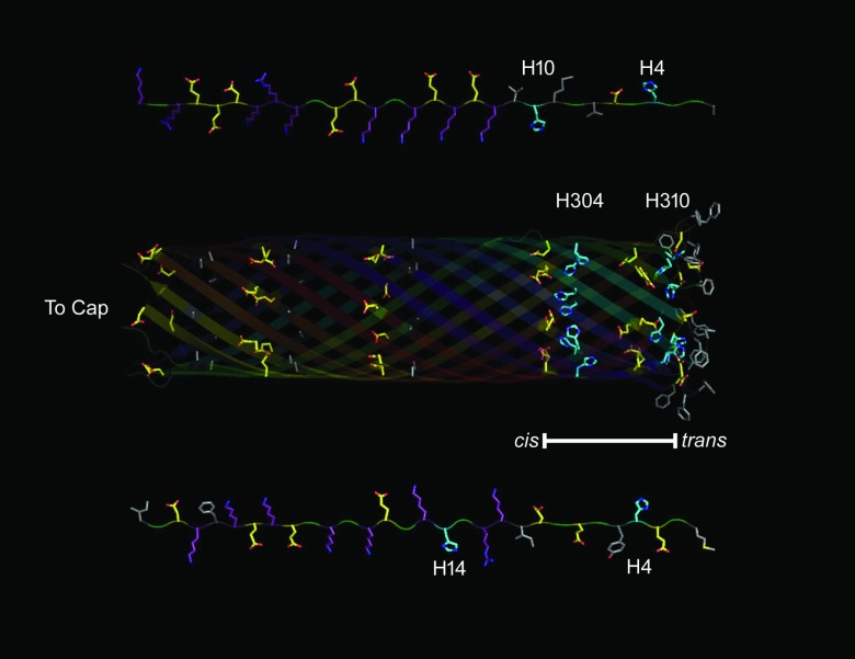
Figure 7.
Protein topology of the disordered N-terminus of EF/LF and the lumen of the PA63 channel β-barrel. (Top) Extended chain representation of the first 30 disordered amino acids (right to left) from the N-terminus of LF. (Middle) The lumen of the β-barrel from a model of its structure. (Bottom) Extended chain representation of the first 30 disordered amino acids (right to left) from the N-terminus of EF. PyMol was used to generate the topological representation based on sequences of EF,124 LF,76 and whole model of (PA63)7.22 Residues are colored by the following classifications: Marine – His, Purple – Basic, Yellow – Acidic, Grey – Hydrophobic. Blue and Red denote N and O moieties, respectively. The bar represents the membrane spanning region of the β-barrel.

