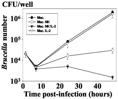
FIG. 2.
Kinetics of the intramacrophagic development of B. suis in the presence of autologous NK cells. Infected macrophages (8 × 10⁵/well) were cultured in 1 ml of culture medium in the presence of NK cells (ratio of NK cells to B. suis-infected cells = 1) under the same conditions as for Fig. 1. In order to assess the role of NK cell activation, rIL-2 was added to the cell culture concomitantly with NK cells at a concentration of 100 U/ml. The number of viable B. suis CFU was then determined at different times p.i., as described in Materials and Methods. Experiments were performed in triplicates and means ± SEMs (error bars) of four different experiments are shown. Symbols: •, infected macrophage alone; ○, infected macrophage incubated with NK cells; ▿, infected macrophages incubated with rIL-2 (100 U/ml); ▾, infected macrophages incubated with NK cells and rIL-2.

