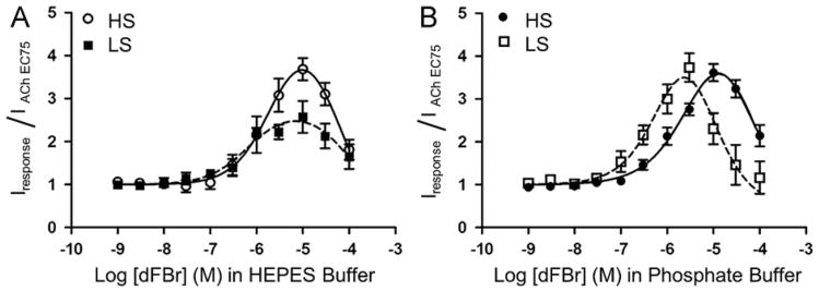
Fig. 5.
Effect of HEPES on potentiation of high sensitivity (HS) and low sensitivity (LS) α4β2 nicotinic acetylcholine receptors by the PAM dFBr. (A) Concentration/response curves obtained from co-application of dFBr and acetylcholine (concentration equal to the acetylcholine EC75) on HS and LS α4β2 nicotinic acetylcholine receptors in HEPES buffer. (B) Concentration/response curves obtained from co-application of dFBr and acetylcholine (concentration equal to the acetylcholine EC75) on HS and LS α4β2 nicotinic acetylcholine receptors in phosphate recording buffer. For all experiments, responses were obtained from Xenopus oocytes under voltage clamp conditions (Vm= −60 mV). Individual peak amplitudes were normalized to the response obtained using an identical concentration of acetylcholine alone (equal to the acetylcholine EC75). pEC50, pIC50 and Imax (%) values (see Table 2) were determined using non-linear curve fitting as described in the methods. Data points represent at least four replicate values obtained from four different oocytes harvested from at least two different frogs. Error bars indicate ± S.E.M.

