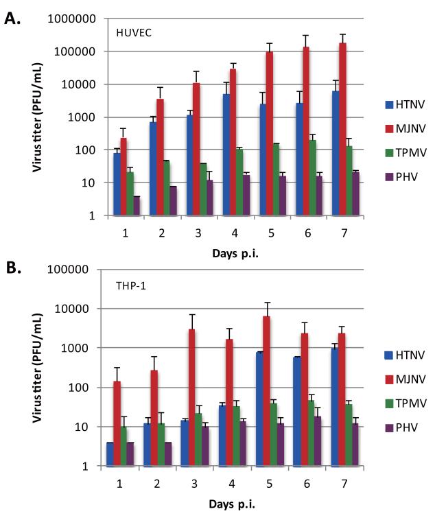
Fig. 2. Hantavirus replication in HUVEC and THP-1 cells.
HUVEC (A) and THP-1 (B) cells were infected with HTNV, MJNV, TPMV or PHV at an MOI of 1.0. Viral titers in the supernatant of infected cells were analyzed 1 to 7 days postinfection by a plaque assay. Days postinfection are indicated on the x axis, and titers are represented as PFU/mL. Error bars represent SD.

