Figure 3.
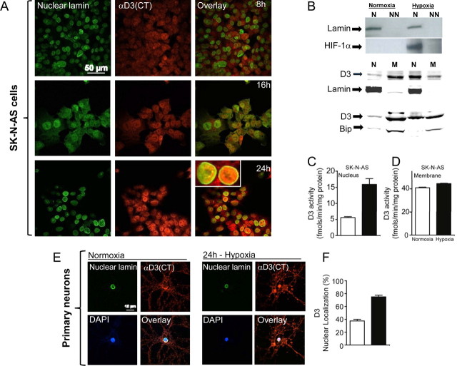
Time-dependent hypoxia-induced nuclear D3 import. A, Immunofluorescence of normoxic SK-N-AS cells processed with α-D3(CT) antibody, α-nuclear lamin antibody, and image overlay. Cells were transferred to a hypoxic chamber (1% O2) and processed for immunofluorescence 8, 16, or 24 h later. B, Western blotting of the indicated SK-N-AS cellular fractions using α-HIF1α, α-nuclear lamin, α-Bip, and α-D3(CT) antibodies; hypoxia lasted for 24 h. N is nucleus (800 g pellet), NN is non-nuclear supernatant prepared in 0.5% Triton X-100 after 800 × g centrifugation; N′ is nucleus (800 g pellet), M is membrane pellet prepared by centrifugation only, after homogenization by ball homogenizer, no detergent was used. α-Nuclear lamin, α-Bip antibodies were used as loading control for nuclear (N′) and membrane (M) fraction, respectively. C, D, D3 activity in nuclear (N′) and membrane (M) fractions prepared by centrifugation only; data are the mean ± SEM; n = 2 per group; experiments were repeated 3 times. E, F, Nuclear localization of D3 under hypoxia in primary hippocampal neuronal cells. Quantification of cells that showed nuclear localization of D3 was done by using a confocal scanning microscope.

