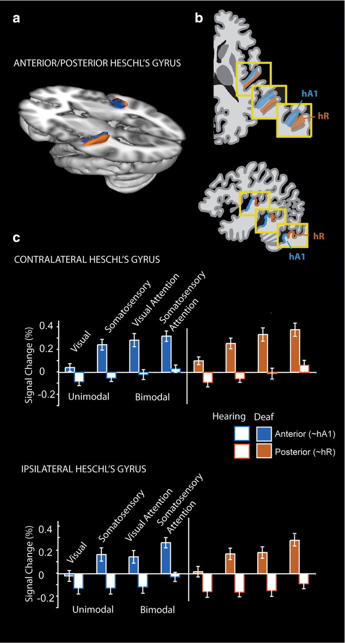
Figure 5.

Anterior to posterior subdivisions of Heschl's gyrus. a, Anatomical Heschl's gyrus ROIs drawn on individual structural brain images were parcellated along the first principle component of voxel centers into an anterior and a posterior subdivision. The divisions are summarized here as a three-dimensional representation at 30% overlap between participants. b, Da Costa et al. (2011) defined human primary auditory cortical areas A1 and R using tonotopy in hearing adults, shown in diagram form here in three examples of anatomical variation. c, Signal change relative to the resting fixation baseline was extracted from individual participant Heschl's gyrus subregions for each block type ipsilateral and contralateral to stimulation for each block type. Deaf participants had larger responses than hearing participants across both regions, and the difference was larger for somatosensory and bimodal stimuli than visual. Error bars represent ± SEM.
