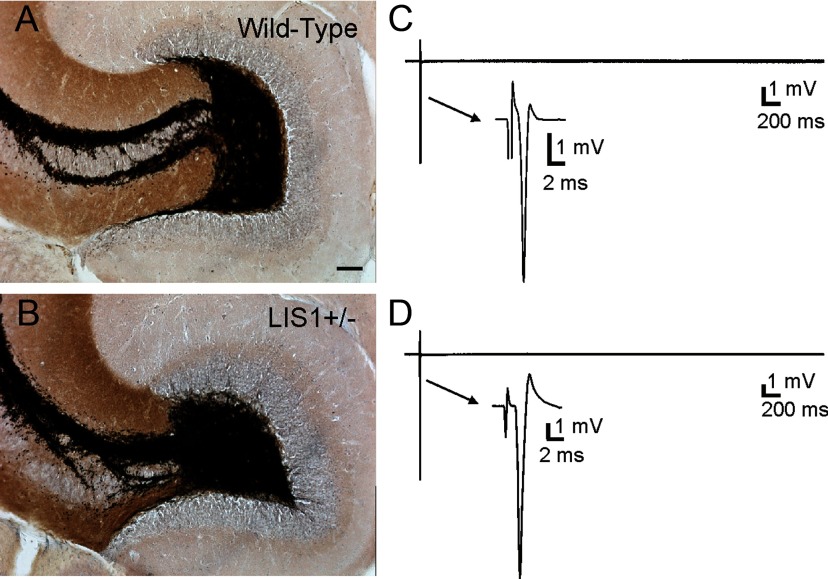
Figure 5.
LIS1 mutant mice do not display abnormal mossy fiber reorganization. A, Timm's- and Nissl-stained section from a wild-type animal. B, Timm's- and Nissl-stained section from an LIS1 mutant mouse. Note the absence of mossy fiber sprouting into the inner molecular layer. C, Hilar stimulation in a wild-type animal evoked a single population spike in Mg2+-free ACSF containing 30 μm BMI. D, Hilar stimulation in LIS1 mutants also evoked a single population spike in Mg2+-free ACSF containing 30 μm BMI. Arrows in C and D indicate expanded portion of the 5-s-long trace to show the population spike. Scale bar, 100 μm.

