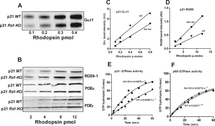
Figure 8.
Quantitative immunoblot analyses (Odyssey imaging system; LI-COR) of key phototransduction protein subunits, transducin α (Gαt1), RGS9, PDE6α, andPDE6γ, in dark-adapted outer segment extracts from P21 WT and Rs1–KO mice (A–D). Transducin α levels relative to rhodopsin were 15–30% lower in Rs1–KO mice than in WT. RGS9, the GAP for Gαt, was 1.7- to 2.5-fold higher in Rs1–KO than in WT. Both PDE6α and PDE6γ protein levels were marginally elevated in Rs1–KO retinas. Replicate gels were run for each antibody tested. The time course of phosphate formation during hydrolysis of [γ-32P]GTP by Gt* in ROS of WT and Rs1–KO mice at P21 (E) and P60 (F). In single-turnover GTPase activity measurements in isolated ROS, GTP hydrolysis by transducin was nearly twofold higher in Rs1–KO than in WT at P21, resulting in a shorter lifetime of activated transducin in Rs1–KO ROS. The rates were not different at P60. The data were fitted to a single-phase exponential decay curve using GraphPad Prism (GraphPad Software).

