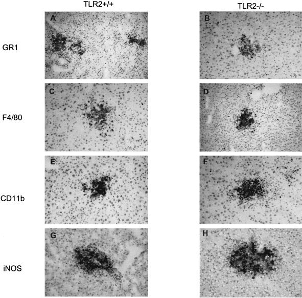
FIG. 7.
Immune cell recruitment to microabscesses after L. monocytogenes infection. Images of the immunohistochemistry of TLR2+/+ (A, C, E, and G) and TLR2−/− (B, D, F, and H) livers 2 days after i.v. infection with 2 × 10⁵ CFU/mouse are shown. Immunolabeling with GR1 (A and B), F4/80 (C and D), CD11b (E and F), and iNOS (G and H) is shown in brown (magnification, ×200).

