Figure 5.
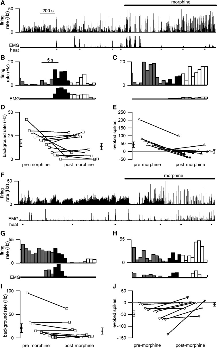
Morphine significantly reduced the responses of ON and OFF cells to noxious stimulation, but had no significant effect on tonic discharge rate. A–C, F–H, Cell discharge before and after morphine (spikes/s, 1 s bins) from representative ON (A–C) and OFF (F–H) cells is shown. Cellular discharge (top trace) and EMG activity (bottom trace) are shown during unstimulated periods and in response to heat stimulation (lines beneath EMG trace). Cells showed significant responses to noxious heat before morphine administration (B, G) but not after morphine (C, H). The time scale in A applies to A and F, and that in B applies to B, C, G, and H. D, E, I, J, For the population of 12 ON (D, E) and 10 OFF (I, J) cells recorded from 18 mice, the average tonic discharge was not affected by morphine (D, I) but the evoked discharge was significantly reduced (E, J). The averages of all values premorphine and postmorphine are shown by the open circles to the left and right, respectively, of the lines. E, J, Open triangles mark cases when a withdrawal was evoked in a majority of the trials analyzed whereas filled symbols mark cases when a withdrawal was not evoked in most trials.

