
Figure 6.
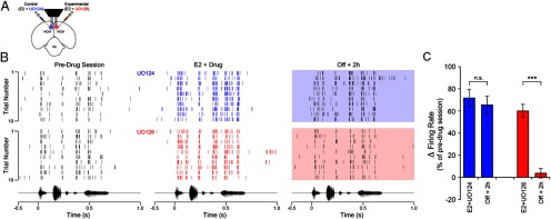
The MAPK-pathway is required for E2-induced, long-lasting enhancement of neurophysiological responses in the awake brain. A, Camera lucida drawing depicting a coronal section through the zebra finch brain illustrating the experimental design for bilateral, multi-electrode neurophysiological recordings coupled to bilateral pharmacological manipulations in awake animals. Animals (n = 44) were repeatedly acclimated to restraint before neural recording sessions, which consisted of a predrug session, a drug session, where animals were continuously infused with a combination of E2 + UO126 (a MEK inhibitor; experimental hemisphere), or E2 + UO124 (the inactive analog of UO126; control hemisphere). After the drug session, infusions were interrupted for both hemispheres, animals were kept in silence for 2 h, and then stimulated with the same song set a final time (Off + 2 h session; see Materials and Methods for details). B, The top panels represent spike raster plots of a representative NCM single-unit before, during (blue), and 2 h after interruption of control infusions (E2 + UO124; shaded blue box). Shown is the spiking behavior of the same neuron across these three recording sessions, during the first 10 renditions of a stimulus. The bottom set of raster plots represent the spiking behavior of another single-unit in NCM recorded simultaneously, in the same animal, in the experimental hemisphere, which received a combination of E2 + UO126. Shown is the response of this representative neuron before, during (red), and 2 h after (shaded red box) interruption of pharmacological infusions. Note that in both control and experimental hemispheres, E2 rapidly enhances the spiking rate of NCM neurons (see E2 + drug sessions). However, although E2 returns to baseline levels in <30 min after interruption of hormonal infusions (Remage-Healey et al., 2010), control hemispheres exhibit a long-lasting increase in the spiking activity of NCM neurons, as previously reported (Tremere and Pinaud, 2011). Remarkably, as a result of the blockade of the MAPK pathway in experimental hemispheres, spiking rates of NCM neurons return to preinfusion levels in the Off + 2 h session; consequently, blockade of the MAPK pathway abolishes this form of plasticity in neural responses induced by E2. The bottom panels represent the amplitude envelope of the stimulus that drove the responses depicted in the raster plots, time aligned with the neural responses. C, Bar graph depicting the variation in the mean firing rate of NCM neurons for “drug” and “Off + 2 h” sessions, for control (blue) and experimental (red) hemispheres. Data are expressed as average percentage changes relative to the pre-drug session. Note that during E2 + UO124 and E2 + UO126 infusions, spiking rates of single-units in NCM significantly increase relative to preinfusion levels. Notably, whereas in control hemispheres the E2-induced increase in discharge rates persists for at least 2 h after interruption of hormonal treatment (see blue Off + 2 h bar), in experimental hemispheres, this long-lasting enhancement of neuronal discharge rates is abolished as a result of blockade of the MAPK pathway with UO126 (see red Off + 2 h bar). (n = 32 neurons/hemisphere). ***p < 0.001.